DVT – Digital Volume Tomography
Digital volume tomography for detailed diagnosis and planning
Three-dimensional x-ray imaging is something most people are familiar with from radiology centers. These computed tomography scans (CT scans) allow for a three-dimensional view of all areas of the body. However, the exposure to radiation this process entails should not be underestimated. CT scans expose patients to some of the highest levels of radiation of any medical diagnostic screening.
We have used CT scans since the mid 1990’s. These images allowed us to present structures of the jaw three-dimensionally prior to placement of an implant. Our patients had to be referred to specialized radiology centers for imaging.
For the last several years, our practice employs the latest in three-dimensional x-ray technology: digital volume tomography (DVT). DVT at our practice exposes the patient to less radiation; furthermore, the technology allows us to x-ray only those regions that are of concern: the jaw areas and maxillary sinuses.
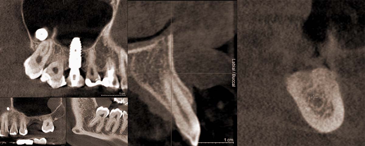
How much bone is available? What is the distance to the nerve canal? In all specialty areas of dentistry, there are many questions that are best answered with 3-D imaging. The third dimension increases clinical safety.
DVT is a three-dimensional imaging tomography process that utilizes x-rays. It is indicated in all cases of extraordinary anatomy or reduced bone substance in the area for placement of the implant. However, we also use digital volume tomography for diagnostic purposes in complex cases and three-dimensional planning on a standard basis. We have been experts in the field of DVT for many years and train our colleagues in specialty courses. We use this technology at our practice to increase the safety of surgical processes and the precision of diagnoses. There are several surgical interventions (immediate placement of implants, navigated implantation) that cannot be performed without DVT. The cumbersome referral to the radiology center is no longer necessary, high exposure to radiation is eliminated, and diagnostics and treatment remain in the same capable hands.
The 3-dimensional images are also helpful to you, the patient, in understanding treatment options. Using the clear images, we can show you the details of your anatomy and provide comprehensive information. It is an important building block in establishing a trusting doctor-patient relationship.
However, such a detailed imaging process is not required for every treatment. The undiscriminating use of radiation is prohibited by law. We therefore strictly adhere to the guidelines of our professional association in determining applicability of the procedure in order to avoid unnecessary exposure to ionized radiation.